A Brief Guide to Partial Necropsy

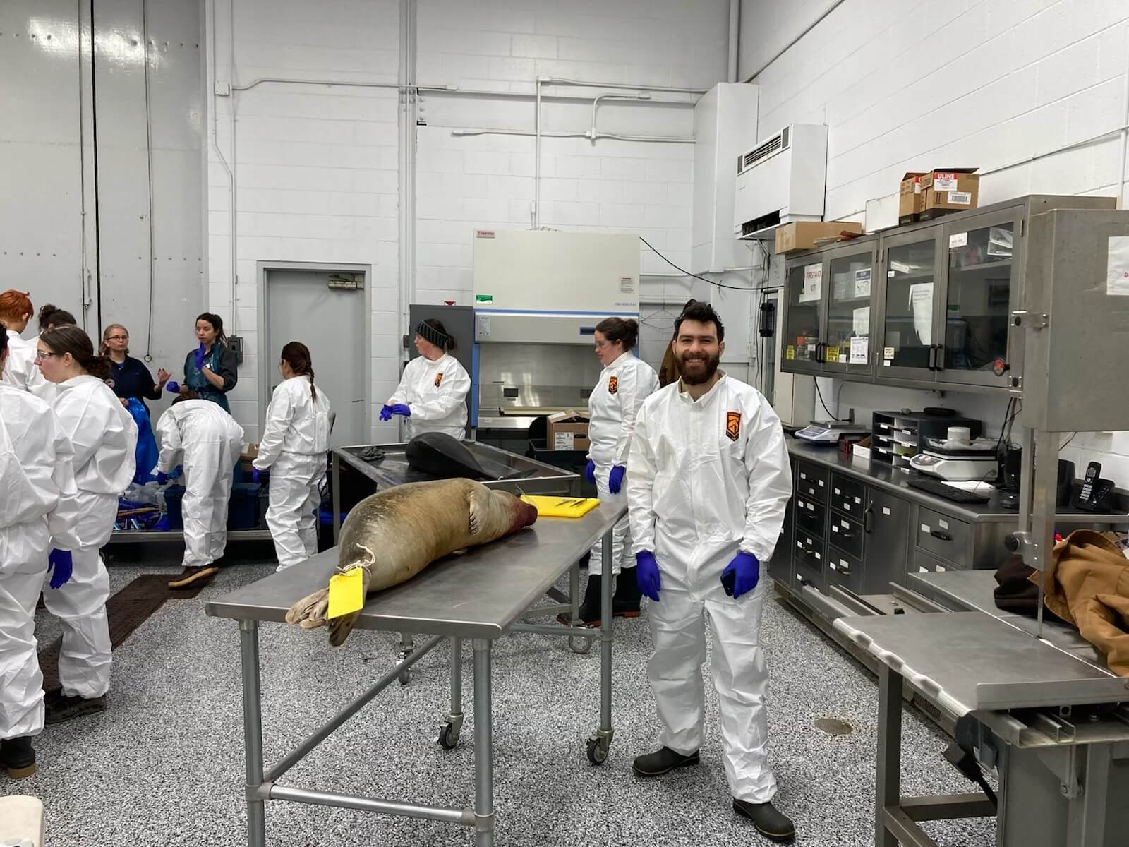
As part of a training course, my colleague and UMM technician Méduline Chailloux and I met on a Saturday morning in a laboratory at the Truro Agricultural Campus in Nova Scotia. Intended for field responders and developed by our colleagues from the Marine Animal Response Society (MARS) and the Atlantic Veterinary College (AVC), this three-day training aims to teach us how to collect the necessary data (in the form of samples and observations) on a carcass to gain insight into the causes of strandings and mortalities. The samples and observations that we collect today from carcasses will then allow the veterinarian-pathologists assigned to these cases to determine the cause(s) of mortality in these individuals.
Why am I telling you about the macroscopic examination of a porpoise carcass? Because Méduline and I are about to dismember and inspect, from the blowhole to the tail, the body of this harbour porpoise to perform a “partial necropsy” of the animal.
This exercise was devised in the event it proves impossible to recover a carcass and the veterinarians are unable to travel to perform a necropsy in the field. Although mobile teams will now be trained to collect more information from carcasses, the expertise of veterinarians is irreplaceable.
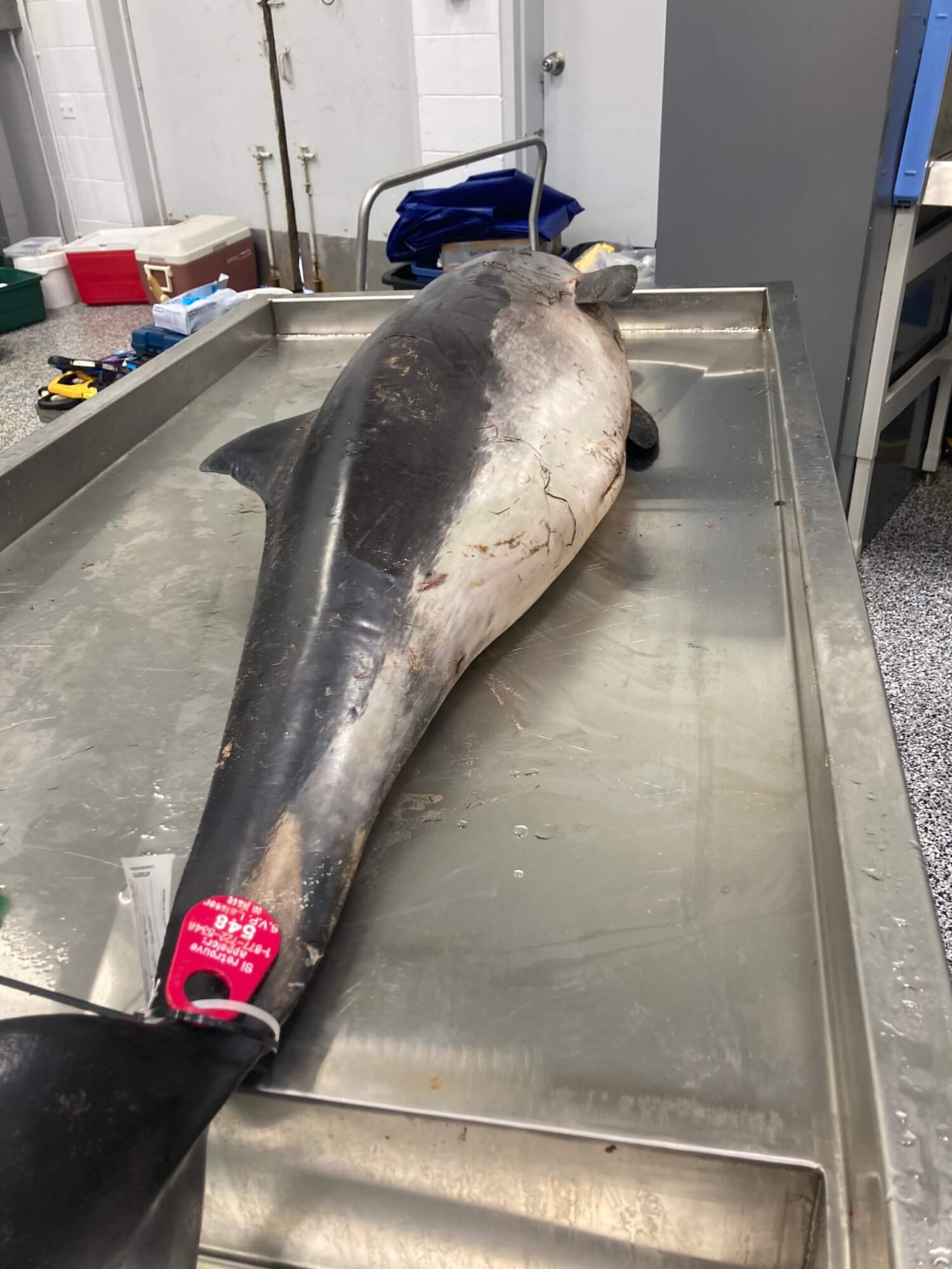
2022-0674 – Adult male harbour porpoise, Magdalen Islands
Carcass reported: October 20, 2022
Interventions carried out:
- Recovery: October 21, 2022 by Satellite Team (IDLM Zip Committee)
- Transport to necropsy site: February 9, 2023 February 9, 2023
- Necropsy: February 25, 2023
Fat thickness: 10 mm
Total length: 155 cm
Girth at anus: 48 cm
Maximum girth: 85 cm
Girth behind flippers: 84 cm
Nutritional condition: slightly emaciated
Apparent lesions: presence of rake marks on the head and left flank, 4 lines, 20 mm long, 5 mm between each line; presence of lesions around the eye
Special observations: no teeth visible in the mouth, no lesions suggesting entanglement, no apparent lesions suggesting blunt trauma
The field is not the lab!
Hydraulic tables, sterile environment, ambient temperature of 21°C, hot water hoses, sinks, carcass winch… These are blissful conditions for collecting samples! On the other hand, I imagine the real-life conditions in which the QMMERN mobile teams will have to take these samples: a sandy beach on the North Shore in October, a northerly wind blowing in your face, an autumn cold snap that numbs your fingertips, blood that gets on anything you handle, sand that fouls exposed animal tissue, changing tides, and the distance you have to travel to the carcass while lugging all your gear with you. We’re a far cry from that here!
Looking for clues
Dressed in our personal protective equipment and equipped with sharp knives, tweezers and surgical scissors, off we go! As we dissect the cetacean, we remain on the lookout for the presence of anything abnormal in the body. These anomalies will guide our sample collection. Our amateur eyes look for any tissue showing signs of bruising, hemorrhages, edema, necrosis, hematomas, hemothorax as well as the presence of any fractures, abnormal masses, signs of malnutrition, scoliosis, parasites, unusual exudates (an organic fluid that oozes from an inflamed surface) and so on and so forth.
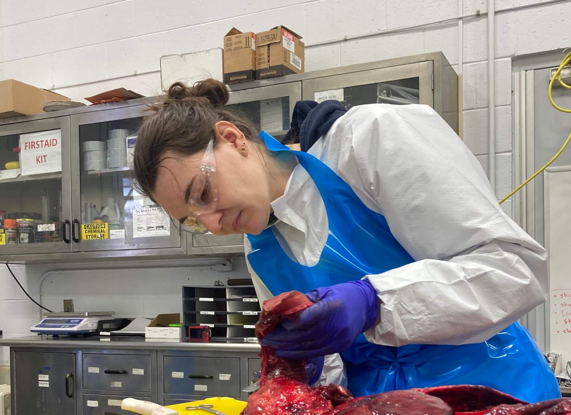
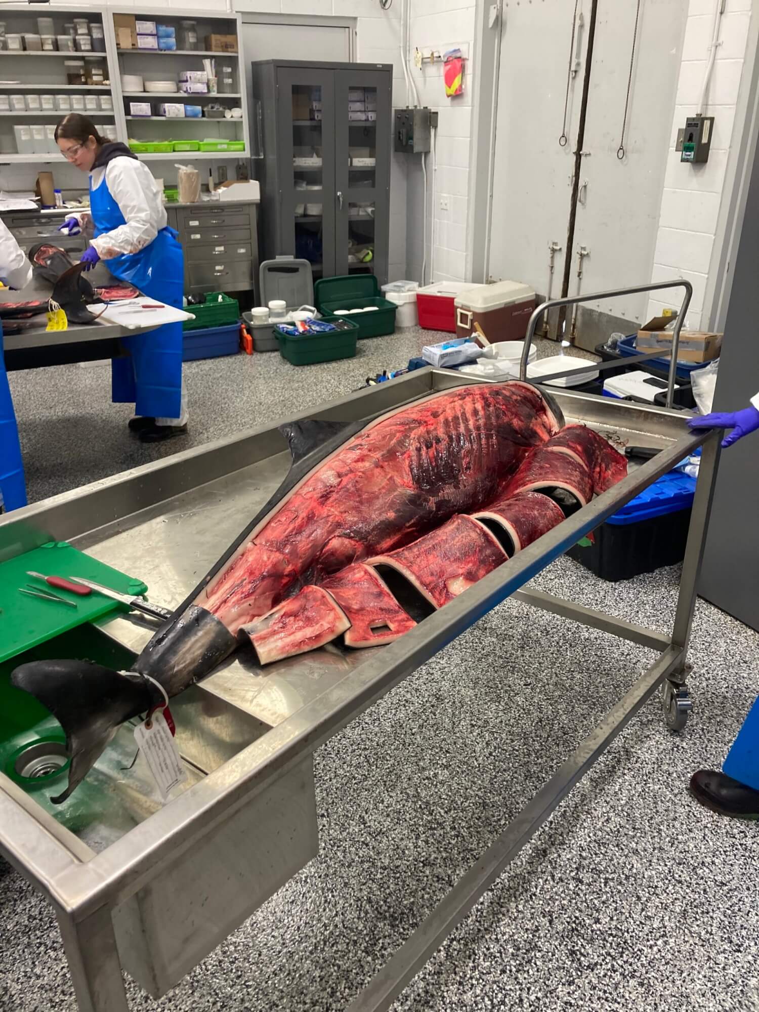
Once the fat and skin are removed from one side of the animal, the rib cage is severed and the internal organs are exposed. It’s a thing of beauty; behind a thick layer of blubber lies an exquisite machine. Laura Bourque, wildlife veterinarian and pathologist from the Atlantic Veterinary College, leads the workshop and describes the impressive morphology of cetaceans. “Everything is designed to facilitate underwater movement while minimizing energy expenditure.” The tight, firm skin of cetaceans is “nearly elastic,” meaning that after each stroke of the caudal fin, the tail returns to its original position without any additional effort. Laura Bourque also points out the characteristics of the animal’s streamlined body, a shape that confers superior hydrodynamics.
Every step of the process is well documented and photographed. The images and notes we take will facilitate a more in-depth analysis later. Each organ is visually analyzed and dissected to reveal lesions and/or signs of disease. Organs are systematically removed, starting with the cardiac and respiratory system: tongue, aorta, trachea, esophagus, heart and lungs. Then we remove the abdominal mass: intestines, stomach, liver and spleen, not to mention the uro-genital system (kidneys, genitalia and adrenal glands). We also take out the muscle mass from the spine to facilitate the search for lesions, in addition to removing the brain from the skull. Lastly, we meticulously dissect each organ to assess its condition while taking samples that will be kept until they are analyzed in the lab by veterinary pathologists.
What does a carcass tell us?
Although veterinary pathologists are able to analyze the tissues we recover to paint a comprehensive picture of the animal, we are still able to extract important tidbits of information during our initial analyses.
For example, the absence of teeth in this porpoise leads us to conclude that it is an older individual. Dissecting the trachea reveals that a large amount of foam has accumulated in the respiratory tract and that significant accumulation is also present in the bronchial tubes of the lungs. “This foam suggests that this porpoise may have drowned,” confirms Laura Bourque. On the other hand, no signs of entanglement were noted during the macroscopic examination. How did it drown? The samples we’ve taken may reveal additional information once we get back to the laboratory. Continuing our exploration toward the gastrointestinal system, we are impressed by the presence in the animal’s stomach of a large colony of nematodes, a type of parasitic worm. “Completely normal,” explains Dr. Laura Bourque. “They can live quite well with large quantities of worms!” With the tissue samples we collect, Dr. Bourque will be able to determine whether certain changes in the animal’s body took place antemortem (before death) or postmortem (after death). Preserved in formaldehyde or stored in a freezer, the tissues taken from the porpoise carcass will also enable Laura Bourque and her team to focus on histology (study of the microscopic structure of the tissues) and thereby confirm the cause(s) of the animal’s death.
I am impressed with the complexity of the work and the effort required to compile important data on marine mammals in the St. Lawrence. Each stakeholder in the process represents an important link in the knowledge acquisition chain. As soon as an incident involving a stranded, drifting or vulnerable marine mammal is reported, the gears of a well-oiled machine are set in motion. Everyone plays their part in obtaining data that will inform major marine wildlife conservation decisions in Canada. We can be proud to be a part of this process as we strive to better understand in order to better protect.
Patrick Weldon
Patrick Weldon joined the GREMM team as UMM supervisor in 2022. He now holds the position of responsible at the Quebec Marine Mammal Emergency Response Network (QMMERN). An anthropologist by training, it is his strong connection with the ocean that sparked his desire to get involved in protecting the marine ecosystem.
Collaboration Spéciale
Recommended articles
25 Years of Evolution and Passion in the Field
It was 1999, and my life was about to change. I had just completed my studies in biology and was…

On board the Bella Desgagnés, sighting of a North Atlantic right whale
By: Valérie Filion head of the interpreting department aboard the Bella Desgagnés for the Marine Mammal Observation Network (MMON) My…
Japan’s Ogasawara Islands: Expedition to Collect Cetacean Sounds and Images
There are many places around the world that lend themselves to watching whales, but some of them are unique. Last…