From Necropsy to Microscope
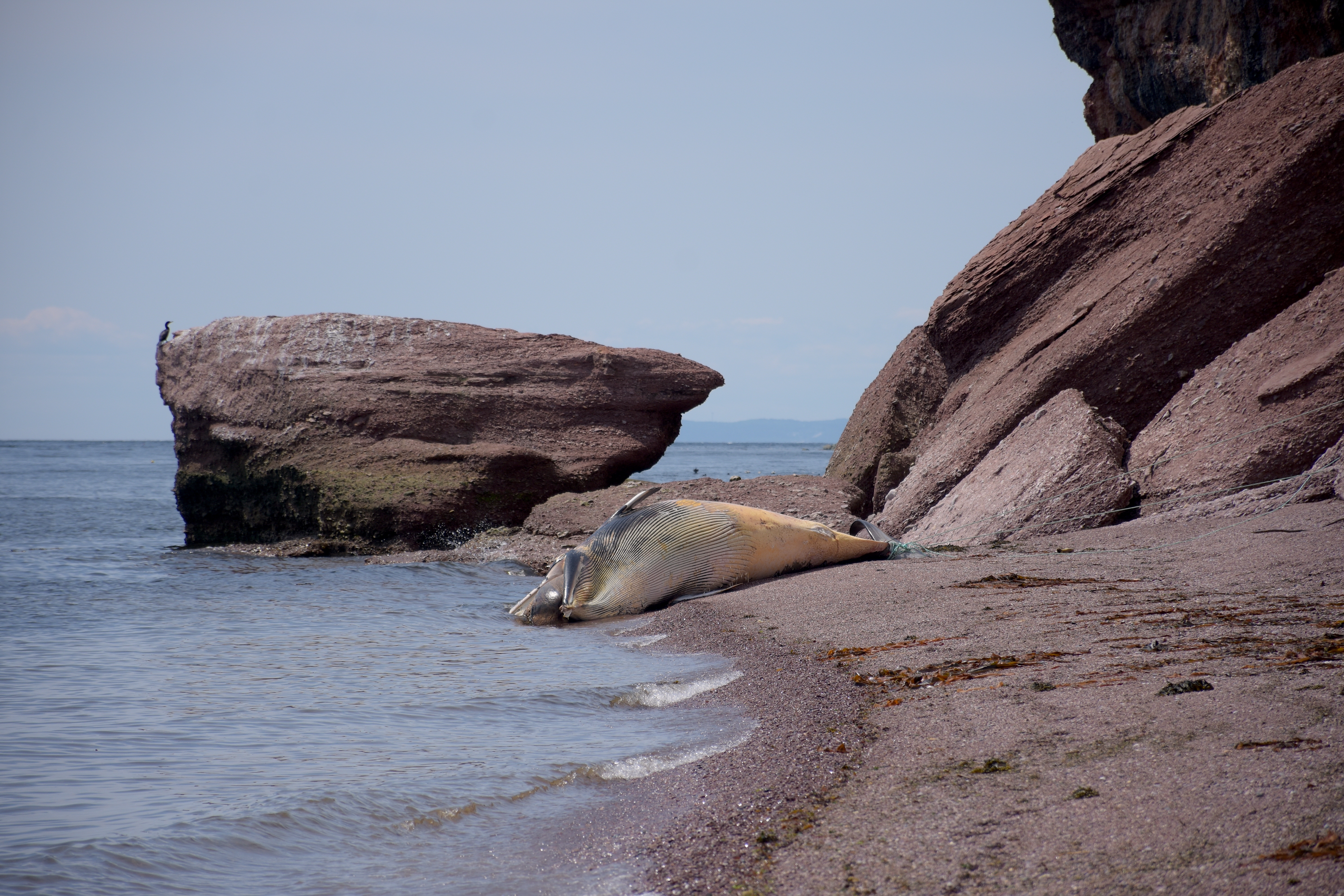
Last week, we tracked the journey of a female beluga from drifting offshore to the Faculty of Veterinary Medicine at the Université de Montréal. This week, discover the necropsy of a male beluga that was discovered on September 10.
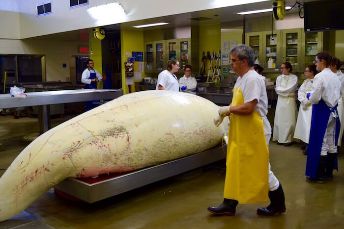
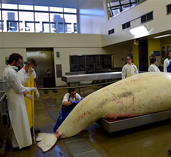
A beluga necropsy is no trivial matter, and this exhaustive examination is is a case in point, as there are over 150 students on a waiting list to participate in the in-depth study of the organs and tissues of a representative of this endangered population. Apprentice veterinarians stand in awe before the lifeless whale, barely contained on the stainless steel table. In a few hours, only a skeleton with its organs exposed will remain. The objective is to shed further light on the health of the St. Lawrence beluga population and to discover the cause of death of this animal, obviously an old specimen judging by its size and worn teeth.
Measurement taking and external photos
The beluga carcass is one of the largest ever studied by the Beluga Carcass Recovery Network since it was first created in 1982. The beluga measures 4.75 m long and weighs 1.3 t. When it was taken out of the freezer where it had been stored overnight, the animal, suspended by the tail, touched the floor, a first for veterinarian Stéphane Lair, who began studying beluga carcasses in the 1980s.
After being careful to respect the numerous rules of hygiene in the room and avoid potential contamination of clothing and accessories, the whale must be measured: total length, half-circumference of the peduncle, stomach, neck, length of the genital slit, thickness of fat. These systematized data are used to establish, among other things, whether or not the animal is in good condition.
The candidate here is quite plump, notably due to the effect of putrefaction gases, but its 10 cm of blubber suggests that it did not die from a long illness but probably from an acute trauma. A sick or old animal that had long stopped feeding would have drawn from its fat reserves and would have been emaciated, which is not at all the case with our beluga.
Photos of the dorsal crest are also taken for submission to the Group for Research and Education on Marine Mammals (GREMM). The GREMM team strives to match photos of carcasses with those of living animals in the photo-ID catalogue. GREMM Scientific Director Robert Michaud would have hoped that the beluga would have evident markings or scars in order to add a chapter to the life of an animal he knows well, but Dr. Lair only notes subtle notches in the dorsal crest. To be continued!
From beluga to samples
Students and veterinarians are busy as bees; first, they remove sections of fat, taking care not to damage the organs that are quickly exposed. Stéphane Lair takes advantage of this moment to teach some basic animal biology. “Did you know that the elephant is the only other mammal besides whales that has internal testicles?” he asks in the large, well lit room, under a putrid, almost unbearable stench.
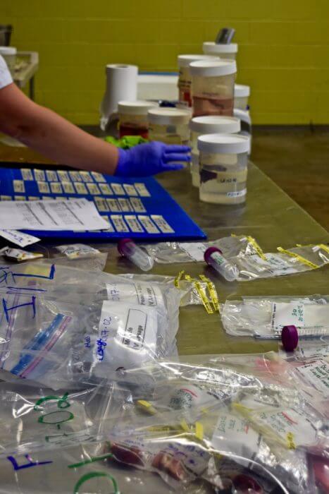
Everything goes quickly and smoothly: the sternum is exposed with the electric winch, the skull is sawn in two to expose the brain, veterinarians are busy unrolling the intestines, removing and laying out the lungs. The stomach contents are quickly packed in a huge ice box that will be sent to the team of Véronique Lesage at the Maurice Lamontagne Institute for study projects. Dozens of jars containing the thyroid gland, heart tissue, lung tissue, liver tissue, etc. are meticulously identified and arranged by Viviane Casaubon, a biology technician and necropsy room attendant at the Centre québécois sur la santé des animaux sauvages (CQSAS). In less than two hours, all traces of the whale are gone, with just samples, a few pieces of skin and liquid left on the floor of the necropsy room.
Animal pathology specialist
Veterinarian Stéphane Lair is experienced: by probing the animal between various mechanical and technical tasks, he has not detected any obvious lesions. At this stage, the cause of mortality of this beluga is unknown. Such is the case for 30% of the belugas studied.
Interested in projects that involve working directly with wildlife, Stéphane Lair has become a leading specialist in wildlife pathologies in Quebec. It was with Dr. Daniel Martineau that he took his first steps. “At the time, we began the necropsy at 10 in the evening and finished at 4 in the morning and then we went for pizza at the 24-hour diner, exhausted,” he remembers. Since they first started, 271 beluga necropsies have been completed out of a total of approximately 560 carcasses found stranded.
Although not all carcasses reveal clear indications of the causes of mortality, as is the case with this sampled male, veterinarian Lair has a very clear picture of the health status of the beluga whale population, the threats to this population and the issues faced by these canaries of the seas.
You can read about the first leg of the journey in the chronicle: From the sea to the necropsy room (1 of 3)
To find out more, watch the video of a beluga necropsy performed at the Faculty of Veterinary Medicine at the Université de Montréal


