From the sea to the lab: the journey of a beluga carcass (3/3)


In the past two weeks, we’ve tracked the journeys of beluga carcasses from the open sea to the Faculty of Veterinary Medicine (FMV) in Saint-Hyacinthe. This week, for the third and final column, discover some of the important issues being raised from the samples taken from these representatives of an endangered population.
From samples to key issues
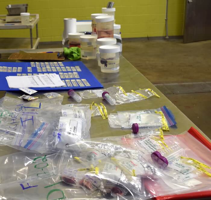
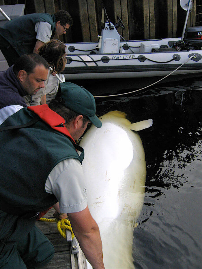
At the end of the necropsy taking place at the Faculty of Veterinary Medicine in Saint-Hyacinthe, just a few bits of tissue and organs remain for further examination. While skin samples will be shipped to Nova Scotia, others will be sent just across the Jacques Cartier Bridge.
Finding the cause
Some belugas have conspicuous lesions that were identified by veterinarians during the necropsy. But in many cases, there is no immediately apparent cause of mortality, hence the importance of histopathological analysis, which entails studying organic tissues under a microscope. The anomalies then detected make it possible to diagnose a pathology or a disease in order to discover the cause of death of the subject.
“For example, if the beluga has a bacterial infection, inflammation will be visible in the tissue,” says Stéphane Lair of the Faculty of Veterinary Medicine. Depending on what is found during the analysis of the tissue slides, the samples will then be submitted to specialized teams at the bacteriology or virology labs.
Dr. Lair and his assistants published results in this regard in 2015. Of the 222 beluga carcasses studied between 1983 and 2012, infectious diseases – which were identified as the leading cause of mortality in the population – were responsible for one-third (32%) of beluga deaths. In juvenile belugas, verminous pneumonia was the cause of death in 52% of the animals. A total of 31 belugas died as a result of infection from malignant tumours, i.e. 20% of belugas aged 19 years or older. One of the hypotheses put forward to explain the unusually high frequency of some of the pathological conditions observed in belugas is the chronic exposure to environmental contaminants.
Heavily contaminated
Based on fat and skin samples, biologist Jonathan Verreault, associate professor at UQAM’s Department of Biological Sciences, is currently developing a portrait of the toxins found in belugas. While the presence of contaminants in beluga tissues has been the subject of long-term investigations, the nature of these contaminants has changed. In the early years of the carcass recovery project, an unusually high number of cancer cases were found in the animals studied. Indeed, St. Lawrence belugas are amongst the most heavily contaminated marine mammals in the world. “When we look at what was present in their environment, the blame often fell on polycyclic aromatic hydrocarbons (PAHs), an important source of which was aluminum production,” explains Dr. Lair. Since the 1970s, PAH concentrations in sediment of the Saguenay have decreased as industrial processes have evolved.
While a number of persistent and toxic chemicals have been banned in Canada, others grew exponentially in the 1990s. Such is notably the case of polybrominated diphenyl ethers (PBDEs), a family of compounds used as flame retardants. PBDE concentrations have increased dramatically in belugas, and these products are known to impact the endocrine system in mammals. Amongst other things, they are believed to have a significant impact on the thyroid gland, especially during calving, which might explain the uptick in observations of newborn carcasses since 2010.
“In addition to the 38 PBDE congeners, we analyze nearly a dozen emerging flame retardants that are substitute compounds for the PBDEs that have been banned in consumer products. We also intend to analyze other emerging compounds and possibly also classical organochlorines such as PCBs, DDTs, HCBs, etc.,” explains Jonathan Verreault.
DNA in the skin
Skin samples are also being sent to the Maritimes. Beluga skin, in addition to contamination-related information, contains valuable data on the whale’s genetic profile. Likewise, if the DNA profile is available, it may be possible to match the carcass to a known living animal.
Whether the carcass is due to undergo a necropsy or not, a skin sample taken from the site of stranding is sent to the lab of Tim Frasier at Nova Scotia’s Saint Mary’s University. This expert geneticist is studies the social structure of the St. Lawrence beluga population.
The fact that the belugas under study show high rates of infection may also be the result of diminished genetic variability, a weakened state of health due to inbreeding, or a combination of these factors.
Pinpointing age by examining the teeth
The beluga’s skull is detached in pieces during the necropsy. The lower jaw is sent to the Maurice Lamontagne Institute of Fisheries and Oceans Canada (DFO). There, lab technician Yves Morin will attempt to determine the age of the animal.
There are many steps and the techniques are constantly evolving. Once the teeth have been extracted and cleaned, an approx. 50 mm thick sliver must be cut lengthwise, polished, and digitized with a high-resolution scanner or a binocular microscope equipped with a digital camera. The photos are then edited to bring out the contrasts in order to better discern the growth rings. A single growth layer represents one year of life. Belugas can live up to sixty years.
Saxitoxin under the researchers’ microscope
Toxic and harmful microalgae are naturally present in the habitat of the St. Lawrence beluga. This phytoplankton varies in abundance from one summer to the next, sometimes creating a “red tide”.
The alga A. tamarense produces a powerful family of so-called paralytic toxins capable of causing a quick death by asphyxiation. In humans, the phenomenon is known as “paralytic shellfish poisoning” (PSP) and is generated by the accumulation of saxitoxin and its derivatives in filter-feeding bivalves. In the case of the beluga, the phenomenon is caused by an accumulation of the toxin in their prey.
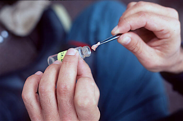
During the necropsy, depending on the animal’s condition, samples from the liver, brain, kidney, urine, blood and digestive tract are set aside to quantify the dosage of the paralyzing toxin. These samples are sent to the laboratory of Michael Scarratt and Sonia Michaud, researcher and biologist, respectively, at DFO’s Maurice Lamontagne Institute.
In recent years, belugas in the Estuary have show signs of chronic exposure. Between 1995 and 2012, 18 of the 66 beluga carcasses tested positive for the presence of saxitoxin and its derivatives in at least one of their tissues. It is difficult to assess whether this contamination is the direct cause of death of the belugas, as there is very little information as to what constitutes a lethal dose. It is possible, however, that in some cases it was an indirect cause.
“If persistent, event low levels of intoxication could influence the animal’s behaviour, weaken it, make it more prone to certain diseases, or more susceptible to stranding or collisions with boats,” suggests Michael Scarratt.
Reconstituting the dinner menu
The biggest container taken out of the necropsy room will also be sent to DFO’s Maurice Lamontagne Institute, this time to Véronique Lesage’s laboratory. A large icebox containing stomach contents will help determine what the beluga consumed before it died.
The contents of the stomach are washed and sieved. Researchers look for prey residues such as squid beaks, crystallized fishbones, otoliths (small crystals from the ears of some fish) and the hooks of polychaetes (bristle worms living in the sediment).
Thanks to this content, researchers can gain insight into the food sources that constitute the diet of the St. Lawrence beluga and thus study the evolution of this aspect of the species’ biology.
Josiane Cabana
Josiane Cabana served as Director for the Quebec Marine Mammal Emergency Response Network call centre from 2011 to 2018. When she’s not responding to cases of dead or vulnerable marine mammals, she likes to take the time to educate local residents on the threats faced by these animals. Biologist by training, she has been involved with the GREMM for more than 15 years, and always with the same undying passion!
Recommended articles
Satellite Teams: Strengthening QMMERN’s Response Capacity across Quebec’s Vast Territory
With fewer phone calls coming into Marine Mammal Emergencies (UMM) in winter than during the high season, it’s the perfect…
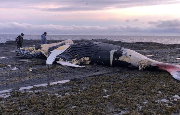
Humpback Observed Earlier This Summer Washes Up in the Gaspé
On Wednesday, October 25, a humpback whale carcass washed up in Saint-Maxime-du-Mont-Louis in the Gaspé Peninsula. It was a male…
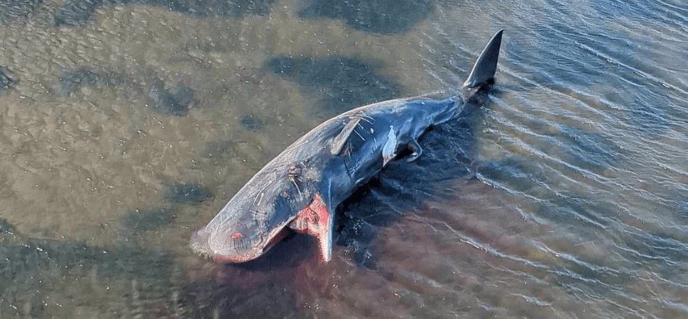
Stranded Sperm Whale on the North Shore
On November 6, flying above the gulf fifteen kilometres from the village of Baie-Johan-Beetz, a helicopter pilot notices a greyish…